Our Doctors

DR. BHARATHI RAJANNA
MBBS, MD OBG, DMAS, ...
| Experience | : | 35 Years Yeras Experience |
|---|---|---|
| Speciality | : | Fertility Specialist... | Location | : | Rajajinagar |
| Timings | : | Mon - Sat : 11:30 AM... |
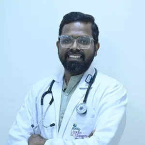
DR. NANDEESH REDDY
MBBS, MS - Obstetric...
| Experience | : | 7 Years Yeras Experience |
|---|---|---|
| Speciality | : | Obstetrics and Gynae... | Location | : | HSR layout 2nd Sector |
| Timings | : | Mon, Wed, Fri & Sat ... |
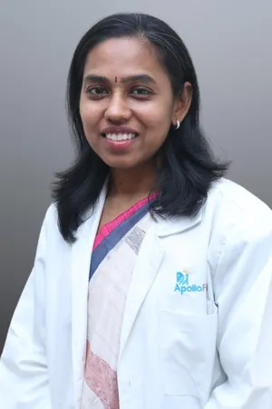
DR. SANGEETHA.S.ANAND
MBBS, MD(OBG), FRM, ...
| Experience | : | 20 Years Yeras Experience |
|---|---|---|
| Speciality | : | Infertility and IVF ... | Location | : | Brookefield |
| Timings | : | Mon to Sat - 09:00 A... |
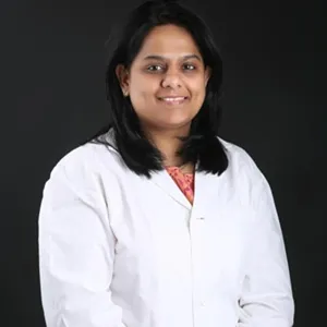
DR. HARSHITA RAMAMURTHY
MBBS, MS (OBG), Fell...
| Experience | : | 15 Years Yeras Experience |
|---|---|---|
| Speciality | : | Obstetrics and Gynae... | Location | : | JP Nagar |
| Timings | : | Mon, Thur & Sat : 8:... |
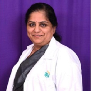
DR. (PROF.) CHITRA RAMAMURTHY
MBBS, MD - Obstetric...
| Experience | : | 37 Years Yeras Experience |
|---|---|---|
| Speciality | : | Obstetrics & Gynaeco... | Location | : | JP Nagar |
| Timings | : | Tues/Thur/Sat : 8.00... |

DR. SANGEETHA S ANAND
MBBS,MD(OBG), FRM, D...
| Experience | : | 20 Years Yeras Experience |
|---|---|---|
| Speciality | : | Fertility... | Location | : | Varthur |
| Timings | : | Mon - Sat : 10:00 AM... |
DR. NIKITHA C P
MS OBG, FMAS, FRM, C...
| Experience | : | 8+ Years Yeras Experience |
|---|---|---|
| Speciality | : | Infertility & Laparo... | Location | : | JP Nagar |
| Timings | : | Mon - Sat : 8:30 AM ... |
DR. KAVITHA V REDDY
MBBS, DGO...
| Experience | : | 20+ Years Yeras Experience |
|---|---|---|
| Speciality | : | IVF Consultant/Gynae... | Location | : | Brookefield |
| Timings | : | Monday to Friday : 0... |
DR. KAVITHA V REDDY
MBBS, DGO...
| Experience | : | 20+ Years Yeras Experience |
|---|---|---|
| Speciality | : | IVF Consultant/Gynae... | Location | : | Brookefield |
| Timings | : | Monday to Friday : 0... |
Ovulation Calculator
![]()